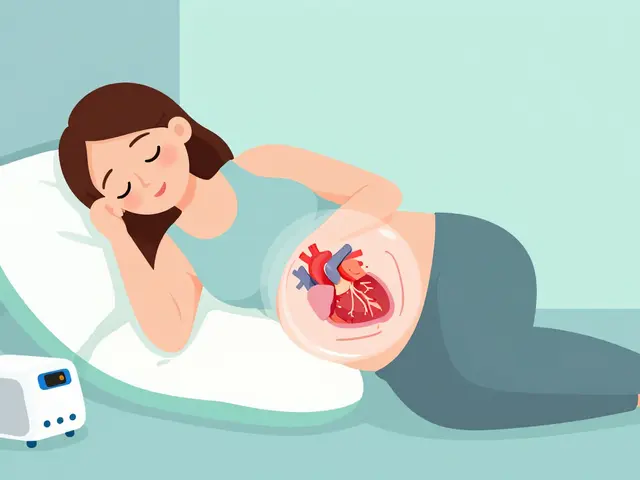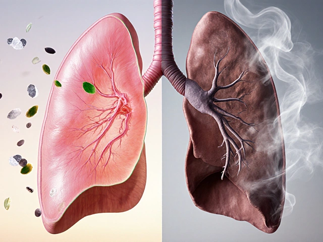
Imagine trying to stand up from a chair or lift your arms to brush your hair, only to find your muscles simply refusing to cooperate. This isn't just fatigue; it’s a signal that something is wrong with the way your body handles inflammation. For thousands of people, this struggle marks the beginning of a journey with Dermatomyositis or Polymyositis, two rare but serious autoimmune conditions. These diseases attack your skeletal muscles, causing progressive weakness that can significantly impact daily life. While there is no cure yet, understanding these conditions and knowing the right treatment path can make the difference between living with disability and regaining your strength.
Understanding the Core Conditions
To grasp what is happening in your body, you first need to distinguish between these two closely related disorders. Both fall under the umbrella of inflammatory myopathies, which means they involve chronic inflammation of the muscles used for voluntary movement. However, they behave differently and require slightly different approaches.
Polymyositis is characterized by symmetrical, progressive weakness in the proximal muscles-those closest to your trunk, such as your hips, thighs, shoulders, and neck. It is a T-cell-mediated disease, meaning your immune system sends cytotoxic T-cells to directly invade and damage muscle fibers. You won’t see skin changes with polymyositis; the battle is entirely internal.
Dermatomyositis, on the other hand, shares the same muscle weakness but adds a distinctive layer of skin involvement. This condition is humorally mediated, involving B-cells and autoantibodies. The hallmark sign is a heliotrope rash-a purple or red discoloration typically appearing on the eyelids, face, neck, elbows, knuckles, and knees. If you have muscle weakness plus this specific rash, dermatomyositis is the likely culprit.
| Feature | Polymyositis | Dermatomyositis |
|---|---|---|
| Skin Involvement | No | Yes (Heliotrope rash, Gottron's papules) |
| Immune Mechanism | T-cell mediated | B-cell/Antibody mediated |
| Cancer Risk Association | Low | Higher (~20% of cases) |
| Lung Involvement | Rare | Common (30-40% develop interstitial lung disease) |
| Pediatric Cases | None reported | Yes (Bimodal age distribution) |
Recognizing the Early Warning Signs
The onset of these conditions is often gradual, which makes early detection tricky. You might notice that climbing stairs feels harder than it used to, or that you’re struggling to rise from a low seat without using your arms. This proximal muscle weakness is the most consistent symptom across both conditions.
For those with dermatomyositis, the visual cues are unmistakable once you know what to look for. The heliotrope rash on the eyelids is often accompanied by swelling. Another key sign is Gottron’s papules-red or purple bumps over the knuckles, elbows, or knees. Beyond the skin and muscles, some patients experience dysphagia, or difficulty swallowing, affecting 15-30% of individuals. This happens because the inflammation spreads to the muscles involved in swallowing.
Fatigue is another massive factor. A 2022 survey by the Myositis Association found that 68% of respondents experienced significant fatigue that limited their daily activities. If you feel exhausted despite adequate rest, and your muscles feel heavy or weak, it’s time to seek medical advice. Don’t dismiss these symptoms as just "getting older" or stress-related burnout.

The Diagnostic Journey: Why It Takes Time
Getting an accurate diagnosis can be frustratingly slow. On average, patients report taking over two years and seeing nearly five specialists before receiving the correct label. Misdiagnosis occurs in about 30% of cases initially, with symptoms often confused with fibromyalgia, lupus, or thyroid disorders.
Here is how the diagnostic process typically unfolds:
- Blood Tests: Doctors will check for elevated creatine phosphokinase (CPK) levels. In active disease, CPK can be 5 to 10 times higher than the normal range (10-120 U/L). They also look for inflammatory markers like ESR and CRP, and specific autoantibodies.
- Electromyography (EMG): This test measures electrical activity in your muscles. In myositis, EMG reveals short-duration, low-amplitude motor unit potentials and spontaneous activity, indicating muscle irritation.
- MRI Scans: Magnetic resonance imaging helps visualize inflammation and damage in specific muscle groups, guiding where to biopsy.
- Muscle Biopsy: This remains the gold standard. A small sample of muscle tissue is examined under a microscope. In polymyositis, doctors look for endomysial inflammatory infiltrates of T cells. In dermatomyositis, they look for perifascicular atrophy and perivascular inflammation.
If you have dermatomyositis, your doctor will likely order a thorough cancer screening. Approximately 20% of dermatomyositis cases are associated with malignancies, particularly ovarian, lung, and gastrointestinal cancers. This step is crucial and distinct from polymyositis management.
First-Line Treatments and Medications
While there is no cure, modern therapy can significantly improve muscle strength and function. The goal is to suppress the overactive immune system to stop the attack on your muscles. Corticosteroids are the cornerstone of initial treatment.
Prednisone is typically started at a high dose, around 1 mg/kg/day (approximately 40-60 mg for an average adult), for 4 to 8 weeks. This aggressive approach aims to quickly halt inflammation. After this initial phase, the dose is gradually tapered down. However, long-term steroid use comes with risks, including osteoporosis, diabetes, and cataracts. About 41% of patients report moderate to severe side effects, with weight gain and insomnia being the most common complaints.
To mitigate these risks and allow for lower steroid doses, doctors often introduce second-line immunosuppressants. Common choices include:
- Methotrexate: Often added if prednisone alone isn’t enough. One patient on a rheumatology forum noted that adding methotrexate reduced their CK levels from 8,200 U/L to 450 U/L within four months, allowing them to taper steroids significantly.
- Azathioprine: Another option for maintaining remission.
- Mycophenolate Mofetil: Frequently used, especially when lung involvement is present.
- Intravenous Immunoglobulin (IVIG): This shows particular efficacy in refractory dermatomyositis cases where other treatments fail.
For stubborn cases, biologics like rituximab have shown promise, with response rates of 60-70% in clinical trials, though they are often used off-label for these conditions.

The Role of Physical Therapy and Lifestyle
Medication stops the fire, but physical therapy rebuilds the house. Starting exercise too early or too aggressively can worsen inflammation, but avoiding movement leads to muscle atrophy. The sweet spot is low-resistance exercise initiated within two weeks of diagnosis.
Tailored exercise programs have been shown to improve functional capacity by 35-45% over six months. Focus on gentle stretching, swimming, or stationary cycling. Avoid heavy lifting until your muscle enzymes (CK levels) normalize and your strength improves. Working with a physical therapist who understands autoimmune myopathies is essential-they can guide you through the Manual Muscle Test (MMT-8) to track progress safely.
Nutrition also plays a role. If you’re on long-term steroids, ensuring adequate calcium and vitamin D intake is critical to protect bone density. Anti-inflammatory diets rich in omega-3 fatty acids may help manage systemic inflammation, though evidence is still evolving.
Prognosis and Future Hope
The outlook for dermatomyositis and polymyositis has improved dramatically. Ten-year survival rates now exceed 80% for dermatomyositis and 85% for polymyositis, a stark contrast to the 50-60% rates seen in the pre-immunosuppressant era. Early, aggressive treatment within the first six months of symptom onset correlates with significantly better long-term outcomes, with 80% of patients achieving remission or low disease activity when treated promptly.
Research is accelerating. Recent trials are investigating JAK inhibitors like tofacitinib, which showed 65% improvement in skin scores for refractory dermatomyositis. New classification criteria incorporating myositis-specific antibodies are expected to reduce diagnostic delays by up to 40%. While challenges remain, the combination of advanced diagnostics, targeted therapies, and supportive care offers real hope for regaining control over your health.
Is dermatomyositis or polymyositis contagious?
No, neither dermatomyositis nor polymyositis is contagious. They are autoimmune diseases, meaning your own immune system mistakenly attacks your body's tissues. You cannot catch them from someone else, nor can you pass them on.
How long does it take to get diagnosed?
Diagnosis can be delayed due to symptom overlap with other conditions. On average, patients report taking 2.3 years and visiting multiple specialists before receiving an accurate diagnosis. Early referral to a rheumatologist can speed up this process.
Can these conditions go into remission?
Yes, many patients achieve remission or low disease activity with timely treatment. Approximately 80% of patients respond well to early aggressive therapy, allowing them to maintain normal daily functions and quality of life.
What is the link between dermatomyositis and cancer?
About 20% of dermatomyositis cases are associated with underlying malignancies, such as ovarian, lung, or gastrointestinal cancers. This association is much weaker or absent in polymyositis. Therefore, comprehensive cancer screening is a standard part of the initial workup for dermatomyositis.
Are there any new treatments available in 2026?
Recent advancements include the use of JAK inhibitors like tofacitinib for refractory dermatomyositis, showing significant improvements in skin and muscle symptoms. Clinical trials are also exploring abatacept for polymyositis. Always consult your rheumatologist about eligibility for these newer therapies.
 Medications