
Most people think of colon polyps as a single thing: a bump on the inside of your bowel that needs to be removed. But if you’ve recently had a colonoscopy report mentioning terms like adenoma is a type of precancerous growth in the colon characterized by abnormal cell structures that can progress to cancer over time, "tubular," or "serrated," you might feel confused. These aren't just different names for the same problem. They are distinct biological entities with different growth patterns, detection challenges, and risks.
Understanding the difference between adenomatous polyps (adenomas) and serrated lesions is crucial because they represent two separate pathways to colorectal cancer. One follows a traditional route we’ve understood for decades; the other is a stealthier, more recent discovery that accounts for a significant portion of modern cases. Knowing which one you have helps determine how closely you need to be monitored and what questions to ask your gastroenterologist.
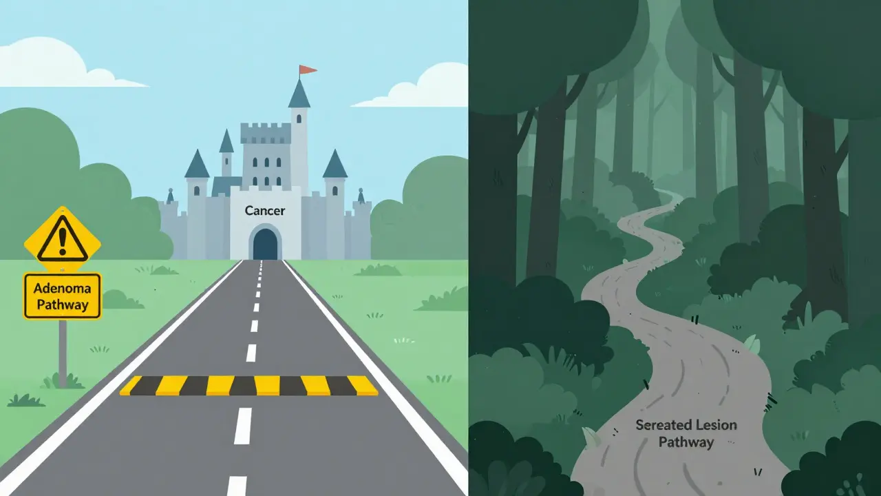
The Two Main Pathways to Colon Cancer
To understand why these polyps matter, we first need to look at how they form. For a long time, doctors believed almost all colon cancers started from conventional adenomas. This is still true for about 70% of cases. However, research has clarified that another group, called serrated lesions, plays a major role in the remaining 20-30% of colon cancers.
Think of it like two different roads leading to the same city. The adenoma road is well-paved and heavily traveled. We know where the speed bumps are (size and shape changes), and we have good signs to warn us. The serrated lesion road is newer, winding through different terrain, and often lacks clear signage until you’re already close to the destination. Both roads lead to cancer, but they require different navigation strategies.
| Feature | Adenomas (Conventional) | Serrated Lesions |
|---|---|---|
| Prevalence | ~70% of all polyps | ~30% of all polyps |
| Molecular Driver | APC gene mutation (Chromosomal Instability) | BRAF mutation (CpG Island Methylator Phenotype) |
| Typical Location | Left side of colon (descending/sigmoid) | Right side of colon (cecum/ascending) |
| Detection Difficulty | Moderate (often pedunculated/stalked) | High (often flat/sessile, easy to miss) |
| Cancer Progression Speed | Slow (10-15 years typically) | Variable, can be faster in some subtypes |
Breaking Down Adenomas: The Classic Precursors
Adenomas are the most common type of precancerous polyp. When your doctor finds an adenoma, they classify it based on its microscopic structure. This classification matters because not all adenomas carry the same risk.
There are three main subtypes:
- Tubular Adenomas: These make up about 70% of all adenomas. They have a tube-like structure under the microscope. While they are precancerous, they tend to grow slowly and have the lowest risk of turning into cancer among adenomas, especially if they are small (less than 1 cm).
- Tubulovillous Adenomas: Accounting for roughly 15% of cases, these are a mix of tubular and villous features. Their risk level sits somewhere in the middle.
- Villous Adenomas: These comprise the remaining 15%. They have finger-like projections and are flatter. Villous components are more aggressive. An adenoma with villous features has a significantly higher chance of containing cancer cells compared to a purely tubular one of the same size.
Size is the other critical factor. An adenoma smaller than 0.5 cm (about half the width of a pencil eraser) has less than a 1% chance of having cancer. However, once an adenoma grows larger than 1 cm, that risk jumps to 10-15%. This is why complete removal during colonoscopy is non-negotiable. If any part remains, it continues to evolve.
Serrated Lesions: The Stealthy Challengers
If adenomas are the classic villains, serrated lesions are the sneaky ones. They get their name from their "saw-tooth" appearance under a microscope. Unlike adenomas, which usually bulge out into the colon, many serrated lesions lie flat against the wall. This makes them notoriously difficult to spot during a standard colonoscopy.
Serrated lesions fall into three categories, each with a very different risk profile:
- Hyperplastic Polyps: These are generally benign, especially when found in the lower (left) part of the colon. Small hyperplastic polyps in this area are often considered low-risk and may not even require immediate removal if they are tiny and clearly identified. However, larger ones or those in the upper colon need attention.
- Sessile Serrated Adenomas/Polyps (SSA/Ps): This is the big concern. SSA/Ps are flat, broad-based, and often located in the right (proximal) colon. A 2016 study published in Colorectal Disease showed that SSA/Ps have a malignant potential equivalent to conventional adenomas, with high-grade dysplasia or carcinoma present in about 13% of cases. Because they are flat, they have a "miss rate" of 2-6% during standard screenings, meaning they can hide in plain sight.
- Traditional Serrated Adenomas (TSAs): These are rarer but carry a high risk of cancer. They often contain BRAF mutations and can progress quickly if not removed completely.
Dr. Matthew Kalady from Ohio State University notes that the serrated pathway accounts for 15-30% of all colorectal cancers. The insidious nature of SSA/Ps lies in their location and shape. They sit quietly in the cecum (the beginning of the large intestine), often without causing symptoms, until they have already progressed.
Why Detection Is Harder Than You Think
You might wonder: "If I had a clean colonoscopy, am I safe?" Not necessarily. The challenge with serrated lesions, particularly SSA/Ps, is detection. Standard colonoscopy relies on visual cues. Pedunculated polyps (those on stalks) stick out and are easy to see. Flat, sessile polyps blend into the surrounding tissue.
This is where technology is stepping in. Recent advances include AI-assisted colonoscopy systems, such as GI Genius, which received FDA approval in 2022. In randomized controlled trials published in The Lancet Gastroenterology & Hepatology, these AI tools demonstrated a 14-18% increase in adenoma detection rates. They act like a second pair of eyes, highlighting subtle texture changes that the human eye might overlook.
Furthermore, the quality of bowel preparation is critical. Residual stool can hide flat lesions. Studies show that poor prep quality increases the miss rate significantly. If you are undergoing screening, ensuring a pristine colon is just as important as the skill of the endoscopist.
Surveillance Intervals: How Often Should You Go Back?
The timing of your next colonoscopy depends entirely on what was found. This is where guidelines diverge slightly, but the general consensus is clear: finding a precancerous polyp means you are at higher risk for future polyps.
For conventional adenomas:
- 1-2 small (<1 cm) tubular adenomas: Return in 7-10 years.
- 3-10 adenomas: Return in 3 years.
- Any adenoma with villous features or >1 cm: Return in 3 years.
For serrated lesions, the rules are stricter due to the higher miss rate and rapid progression potential:
- Small (<1 cm) SSA/Ps without dysplasia: Many experts recommend surveillance in 3 years, though some European guidelines suggest 5 years.
- SSA/Ps ≥10 mm or with dysplasia: Surveillance in 3 years is standard per US Multi-Society Task Force guidelines.
- Multiple serrated polyps: Closer monitoring, often 3 years.
It’s important to note that while these intervals seem frequent, they are designed to catch new growths before they become dangerous. As Dr. Elizabeth Platz from Johns Hopkins points out, most patients with these polyps never develop cancer precisely because they are caught and removed early. The surveillance is the safety net.

Molecular Differences: APC vs. BRAF
Under the hood, these polyps operate on different genetic circuits. Conventional adenomas typically follow the chromosomal instability pathway, driven by mutations in the APC gene. This is the classic "gatekeeper" gene that controls cell growth. When it breaks, cells start dividing uncontrollably.
In contrast, serrated lesions, especially SSA/Ps, often follow the CpG island methylator phenotype (CIMP) pathway. Here, the key player is the BRAF gene. Instead of breaking a gatekeeper, the cell silences tumor suppressor genes through methylation (adding chemical tags to DNA). This epigenetic change leads to uncontrolled growth. Understanding this distinction is becoming increasingly important for personalized medicine. Future treatments may target these specific molecular pathways rather than treating all polyps the same way.
What You Can Do Now
If your pathology report mentions either adenomas or serrated lesions, don’t panic. These findings are actually a success story-they were found before they became cancer. Here are practical steps to take:
- Confirm Complete Removal: Ask your doctor if the polyp was removed entirely. Fragmented removal might require a repeat procedure sooner.
- Clarify the Subtype: Know exactly what type you have. Is it a tubular adenoma or a sessile serrated lesion? This dictates your next appointment date.
- Discuss AI-Assisted Screening: If you are due for a follow-up, ask if your facility uses AI-enhanced colonoscopy or chromoendoscopy (dye spraying) to improve detection of flat lesions.
- Optimize Bowel Prep: Follow prep instructions meticulously. A clean colon is essential for spotting subtle serrated lesions.
- Consider Family History: If multiple family members have had serrated lesions or early-onset colon cancer, discuss genetic counseling. Lynch syndrome and other hereditary conditions can influence polyp types.
Remember, the goal of screening isn't just to find cancer; it's to find the polyps that could become cancer. By understanding the difference between adenomas and serrated lesions, you empower yourself to advocate for the right level of care and surveillance.
Are serrated polyps more dangerous than adenomas?
Not necessarily more dangerous in terms of final outcome if detected and removed, but they are harder to detect. Sessile serrated lesions (SSA/Ps) are flat and easily missed during colonoscopy, leading to a higher "miss rate." Once found, both adenomas and SSA/Ps have similar malignant potentials (around 13% risk of high-grade dysplasia or cancer). The danger lies in the detection gap, not inherently worse biology.
How soon after removing a serrated lesion should I have another colonoscopy?
Guidelines vary slightly, but generally, if you have a sessile serrated lesion (SSA/P) that is 10mm or larger, or has dysplasia, you should return for a surveillance colonoscopy in 3 years. Smaller SSA/Ps without dysplasia might allow for a 3-5 year interval depending on your doctor's assessment and regional guidelines. Always follow the specific recommendation provided by your gastroenterologist based on your full pathology report.
Can lifestyle changes prevent the formation of these polyps?
While genetics play a role, lifestyle factors significantly influence risk. Maintaining a healthy weight, exercising regularly, limiting red and processed meats, and avoiding smoking and excessive alcohol consumption are proven to reduce the risk of developing both adenomas and serrated lesions. Some studies also suggest that adequate calcium and fiber intake may offer protective benefits.
What is the difference between a hyperplastic polyp and a sessile serrated adenoma?
Both are types of serrated polyps, but their risk levels differ drastically. Hyperplastic polyps, especially small ones in the left colon, are generally benign and have negligible cancer risk. Sessile serrated adenomas/polyps (SSA/Ps) are precancerous and can progress to cancer via the serrated pathway. Distinguishing between them requires careful microscopic examination by a pathologist, as they can look similar grossly.
Does AI really help find more polyps?
Yes. Clinical trials have shown that AI-assisted colonoscopy systems can increase adenoma detection rates by 14-18%. These systems analyze the video feed in real-time and alert the endoscopist to potential lesions, particularly flat or subtle ones that might otherwise be overlooked. This is especially valuable for detecting serrated lesions, which are naturally harder to see.
 Medications


Hassan Bukhari
June 6, 2026 AT 06:03It is genuinely exhausting to watch people panic over basic gastroenterology reports without understanding the underlying pathology. The distinction between adenomatous and serrated pathways is not rocket science, yet here we are treating it like state secrets. If you cannot distinguish between a tubular adenoma and a hyperplastic polyp, perhaps you should reconsider your medical literacy before offering unsolicited advice on surveillance intervals.
Aishwarya Thankachan
June 7, 2026 AT 14:31OMG this is so scary 😱 I had a colonoscopy last year and they found something but I didn't ask what kind because I was too scared to look at the report 📄😰 Now I'm wondering if my doctor missed a sessile serrated lesion because they are flat and sneaky 😨 BRAF mutations sound so aggressive 💀 Should I call them immediately or wait? My anxiety is through the roof right now 📈🤯
Jerry Mathews
June 7, 2026 AT 15:56Hey there, take a deep breath. It's completely normal to feel overwhelmed by medical jargon, especially when it involves cancer risks. The good news is that modern screening is incredibly effective at catching these issues early. Whether it's an adenoma or a serrated lesion, the fact that it was found means you're already ahead of the game. Trust your gastroenterologist; they are trained to handle these distinctions with precision. You've got this, and staying informed is a great first step toward peace of mind.
Aswin Narayan J
June 8, 2026 AT 04:18In India, we see a massive surge in colorectal cancer cases among younger demographics, largely due to dietary shifts and sedentary lifestyles. This article highlights a critical gap in global awareness: the stealthy nature of serrated lesions. While Western medicine focuses heavily on APC gene mutations, we must acknowledge that environmental factors play a huge role in triggering these pathways. Ignoring the cultural context of diet and genetics is a mistake many international health organizations make. We need more localized data, not just generic guidelines from the West.
Jennifer Legore
June 8, 2026 AT 05:47I am absolutely thrilled to see such a comprehensive breakdown of these complex topics! :) It is truly wonderful how technology, like AI-assisted colonoscopy systems, is enhancing our ability to detect these subtle lesions. The 14-18% increase in detection rates mentioned in the post is a fantastic statistic that gives me hope for better outcomes globally. Let us all embrace these advancements with open arms and encourage our healthcare providers to adopt the latest tools for optimal patient care. Together, we can make a significant difference! :)
Alyssa Zucker
June 9, 2026 AT 18:04I read through this quietly and felt a bit uneasy. The part about flat lesions hiding in plain sight really stuck with me. I never thought about how the shape of the polyp affects whether it gets seen or not. It makes me wonder if my previous clean bill of health was thorough enough. I don't want to alarm anyone, but it does make you think twice about trusting the process blindly.
Francis Saul
June 9, 2026 AT 18:34look i get why ppl r worried but dont stress urself out. most of these things r caught early and fixed easy. just make sure u drink the prep stuff all the way down so the doc can see everything. if u miss spots its harder to find the flat ones. keep it simple and follow up when told. no biggie really.
Dave Villeneue
June 10, 2026 AT 03:19The reliance on visual inspection during standard colonoscopy is fundamentally flawed. The miss rate of 2-6% for SSA/Ps is unacceptable in a high-stakes diagnostic environment. Physicians who fail to utilize chromoendoscopy or virtual chromoendoscopy are practicing negligence. The data clearly indicates that traditional white-light endoscopy is insufficient for detecting sessile serrated lesions. Immediate adoption of advanced imaging protocols is mandatory, not optional.
Rachel Harrypersad
June 10, 2026 AT 09:27we chase shadows while the real monster hides in the cecum. the body is a labyrinth of deceit. serrated lesions mock our linear thinking. we think straight lines lead to truth but cancer curves around our expectations. why do we fear the unknown so much when it is already inside us. silence is loud in the colon.
Brian Irwin
June 12, 2026 AT 04:40i hear what everyone is saying and it's a lot to take in. honestly just doing the prep right is half the battle. if the colon isn't clean nothing else matters. i know it sucks drinking that stuff but trust me it saves lives. don't let the fancy terms scare you off just show up and do the work. you'll be fine.
Rosy Centire
June 12, 2026 AT 06:10Let us be unequivocally clear: the classification of polyps is not merely academic; it is clinically imperative. The assertion that hyperplastic polyps in the rectosigmoid colon are benign is accurate, yet the extrapolation of this safety to proximal lesions is a dangerous fallacy. Sessile serrated adenomas/polyps (SSA/Ps) require rigorous excision and meticulous surveillance. Any physician who dismisses the significance of BRAF mutations and CpG Island Methylator Phenotype is failing their ethical duty. Precision in terminology saves lives.
Aswin Ashokan
June 12, 2026 AT 16:55another western obsession with over-diagnosing. in my country we deal with real problems not tiny bumps in the gut. this article is full of unnecessary complexity designed to sell more tests. ignore the hype. eat less processed food and stop worrying about every little cell mutation. simplicity works better than expensive machines.
William Storm
June 13, 2026 AT 12:36One might ponder the existential dread induced by the mere possibility of a flat lesion evading detection. Is our existence defined by the unseen threats lurking within our own biology? The pretension of medical authority often masks the sheer randomness of cellular decay. We build elaborate theories-APC genes, BRAF mutations-to impose order on chaos. Yet, the colon remains a dark mirror reflecting our vulnerability. Perhaps the true pathology lies in our need to control the uncontrollable.
Wendy Engelmann
June 13, 2026 AT 17:38I sat back and watched the thread unfold. It's interesting how fear drives so much of the conversation here. The science is solid, but the emotional reaction varies wildly. Some see a threat, others see a puzzle. I prefer to observe rather than intervene. The information is out there for those who seek it calmly. No need to rush into conclusions or panic. Just breathe and let the facts settle.